Rhinoplasty (nose surgery) is one of the most sophisticated procedures in plastic surgery, both in terms of surgical technique and artistic perspective. A successful operation aims to create a nose that is in harmony with the facial features, looks natural, and—most importantly—allows the patient to breathe comfortably. However, due to the dynamic nature of nasal tissue, individual variations in the healing process, and sometimes technical limitations encountered during the initial surgery, the desired outcome may not always be achieved after the first (primary) surgery.
This is where revision rhinoplasty (secondary rhinoplasty) comes into play. Revision rhinoplasty refers to corrective surgeries performed to correct aesthetic deformities that arise after the initial surgery or to restore lost nasal functions (such as breathing).
So, how do you know if everything is going well after your first surgery? How do you distinguish between normal swelling during the healing process and a genuine need for revision? In this comprehensive guide, we’ll detail the 9 early signs that may indicate a need for revision, the anatomy of the healing process, and how to take the right steps at the right time.
The Anatomy of Post-Rhinoplasty Healing: Is It Swelling or a Permanent Problem?
When you look in the mirror after rhinoplasty, the first image you see is not your final result. The nose is one of the organs in our body that retains swelling the longest and has the slowest healing process. For this reason, it is crucial to understandthe “normal healing timeline” before assessing signs that a revision may be needed.
First 3 Months: There is significant swelling along the bridge and tip of the nose. During this period, the nose may appear firm, asymmetrical, and more elevated than usual. Most of these asymmetries result from the swelling not being distributed evenly on both sides.
Month 6: Approximately 70–80% of the swelling subsides. The contours of the nose begin to become more defined, though the tip of the nose may still retain some firmness and thickness.
1st Year (Final Result): The soft tissues heal completely, and the cartilage and bone structures settle into their new form. In patients with thick skin, this process may take up to 1.5–2 years.
Key Rule: Not every instance of asymmetry or shortness of breath observed in the early stages indicates a need for revision surgery. For a true revision assessment, at least one full year must have passed since the surgery. However, the following nine early signs are early indicators that a corrective surgery may be needed at the end of this process.
9 Early Signs That May Indicate the Need for Revision Rhinoplasty
As you monitor the healing process of your nose in the months following surgery, if the following symptoms become permanent or become more pronounced over time, this may indicate the need for a secondary procedure:
1. Breathing Problems That Persist or Worsen After Surgery
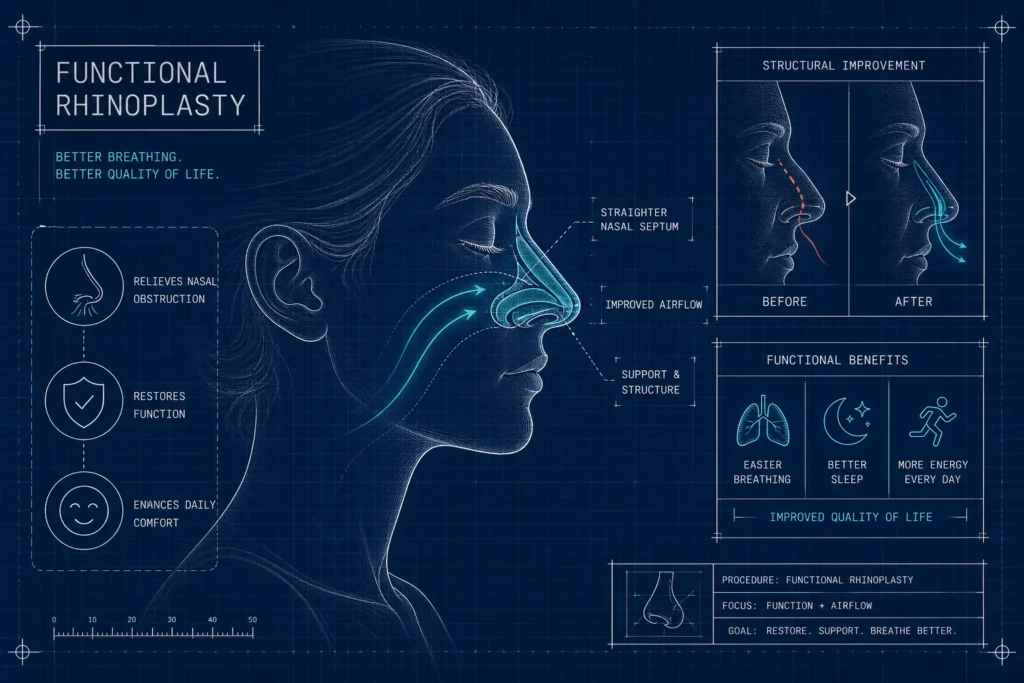
The primary goal of rhinoplasty is to create a functionally healthy nose. It is normal to experience difficulty breathing during the first few months after surgery due to swelling and crusting of the nasal mucosa. However, if breathing does not improve after the sixth month or if no air is passing through one nostril, this may indicate a functional failure.
Causes: Failure to fully correct a deviated septum (cartilage curvature) during the initial surgery, valve collapse (nasal valve insufficiency) resulting from excessive narrowing of the nasal roof, or postoperative adhesions (synechiae).
2. Deformities and Asymmetries on the Bridge of the Nose (Pollybeak Deformity and “Twisted Nose”)
As the bridge of the nose (profile) heals, it should form a straight, slightly curved, or masculine contour. In some cases, as swelling subsides, unexpected curves or protrusions may appear on the bridge of the nose.
Pollybeak Deformity: This condition occurs when the area immediately above the tip of the nose (supratip) is higher than the tip itself, resulting in an appearance that resembles a parrot’s beak when viewed in profile. It is caused by leaving too much of the cartilaginous roof in place or by excessive accumulation of scar tissue in that area.
C or S Curvature: If the nasal bone structure cannot be fully aligned with the midline, or as a result of asymmetrical bone shifts during the healing process, the nose may appear curved in a C or S shape when viewed from the front.
3. Excessive Drooping of the Tip of the Nose (Loss of Projection) or Excessive Upturn (“Pigs Nose”)
The height and angle of the tip of the nose determine the overall harmony of the face’s profile.
Nose Tip Drooping: The tip of the nose, which looks very nice immediately after surgery, may begin to droop downward as months pass and due to the effects of gravity. This condition is usually caused by insufficient cartilage grafts (support structures) used to support the nose tip during the initial surgery.
Excessive Protrusion: This condition occurs when the angle between the tip of the nose and the lip (the nasolabial angle) exceeds the ideal range (95–105° for women, 90–95° for men) and the nostrils appear overly prominent when viewed from the front. Commonly referred to as a “pig nose,” this appearance results from aggressive shortening of the nasal bridge and cartilages.
4. Indentation on the Bridge of the Nose or an Excessively Fleshy Appearance in the “Supratip” Region
If the nasal hump is filed down or excised (removed) excessively during the initial surgery, it can cause the nasal bridge to curve inward too much, leading to a “saddle nose” deformity. This condition not only creates a sunken appearance but also narrows the nasal airway, making breathing significantly more difficult.
5. Asymmetry, Collapse, or “Alar Retraction” of the Nostrils
This condition occurs when, when viewed from the front, one nostril is noticeably different in size, height, or shape from the other. As the tissue heals after surgery, one side of the nose may collapse inward (alar collapse), causing that side to stick to the other during breathing. This is both an aesthetic imbalance and a serious functional problem.
6. Inverted-V Deformity When Viewed from the Front
This is a rupture in the middle septum, where the bony and cartilaginous structures of the nose meet. During surgery, after the nasal bridge is removed, the cartilages collapse inward due to insufficient support of the middle cartilaginous septum (failure to use a spreader graft). When viewed from the front, an inverted “V” shape appears in the center of the nose. This is clear evidence that the middle portion of the nasal passage has collapsed.
7. Prominent Cartilage at the Tip of the Nose, Sharp Edges, and Thinning of the Skin
In rhinoplasty, the edges of the cartilage grafts placed to reshape the tip of the nose are not visible during the first few months after surgery due to a thick layer of edema. However, as the swelling subsides over time and the nasal skin thins, the sharp edges of these cartilage grafts may become visible beneath the skin as asymmetrical protrusions, lumps, or white patches. As the healing process continues, these sharp cartilage edges may irritate the nasal skin from the inside, causing it to thin even further.
8. Permanent Redness and Skin Tissue Abnormalities on the Bridge or Tip of the Nose
The skin of the nose and the underlying soft tissue are affected by the dissection (separation of tissues) performed during surgery. If the subcutaneous tissues were thinned too aggressively during the initial surgery or if blood circulation in the skin was compromised, permanent redness at the tip of the nose, broken capillaries, or hard, irregular scar tissue (fibrosis) may develop during the healing period.
9. Psychological Unhappiness and Aesthetic Dissatisfaction Despite Realistic Expectations
Sometimes the nose may function perfectly well for breathing and have no major anatomical defects. However, the resulting new nose may be completely out of character with the patient’s facial features, ethnic background, or personal identity. If, at the end of one year, the patient—even when viewing the reflection in the mirror objectively and realistically—cannot recognize the image as their own face, and this situation negatively affects their social or psychological well-being, this is also a valid reason for revision surgery.

When Is the Right Time for Revision Rhinoplasty?
If you notice one or more of the above signs in the early stages, you shouldn’t panic. It takes time for the tissues to mature, for the hard areas to soften, and for microcirculation (blood flow) to return to normal.
An early revision procedure performed during an incomplete healing process may lead to further tissue damage and scar formation rather than resolving the existing problem. Therefore, the generally accepted rule for revision surgery is that at least 12 months (ideally 18 months) must have passed since the initial surgery.
However, in certain exceptional cases (such as a very sharp bony protrusion on the bridge of the nose that poses a risk of skin perforation, or acute airway obstruction that completely paralyzes the patient), an earlier procedure may be scheduled at your surgeon’s discretion.
Literature and Scientific Approaches in Secondary Surgery
Revision rhinoplasty is not merely a matter of addressing aesthetic concerns, but also a complex form of tissue engineering. A review of cases involving secondary rhinoplasty in the medical literature reveals that the time elapsed since the initial surgery and accurate tissue analysis directly influence the success of the procedure. Scientific studies published in the world’s most prestigious medical databases emphasize the importance of grafting techniques (cartilage transfer) and careful postoperative follow-up to restore anatomical integrity in revision surgery. If you wish to explore international surgical approaches and case analyses on this topic, you can review current clinical research on revision rhinoplasty through PubMed / National Institutes of Health, a biomedical library containing peer-reviewed medical publications from around the world.
The Importance of Choosing a Surgeon for Revision Surgery and Cartilage Grafts
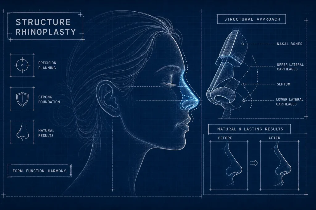
Secondary (revision) rhinoplasty requires a much higher level of surgical experience, anatomical knowledge, and foresight than primary surgery. This is because in revision surgeries, the surgeon is faced with a nose whose anatomy has been disrupted, covered in scar tissue, and—most importantly— whose natural cartilage reserve (septal cartilage) was either used up or damaged during the initial surgery.
Is Rib or Ear Cartilage (Costal and Conchal Graft) Necessary?
To repair a collapsed nasal roof, lift a drooping nasal tip, or correct an inverted V deformity, the surgeon needs new cartilage tissue. When there is insufficient cartilage remaining inside the nose, cartilage is harvested from other areas of the body:
Ear Cartilage (Conchal Graft): It is typically used for tip reshaping and to correct mild depressions. It does not cause any distortion of the ear’s shape.
Rib Cartilage (Costal Graft): This is the gold standard in major revision surgeries where the nasal bridge has completely collapsed (saddle nose) or where very strong structural support is required. It is harvested from the lower chest through a small incision and meticulously sculpted by the surgeon to millimeter precision to form the nose’s new framework.
For this reason, the most important factor determining the success of the surgery is for patients considering revision rhinoplasty to proceed with a plastic surgeon who specializes specifically in this field and has a high success rate and a broad portfolio of revision cases.
Conclusion: It Is Possible to Make Peace with Your Nose Again
Failing to achieve your desired result during your first surgery should not lead you to feel discouraged. Remember that revision rates following rhinoplasty range from 5% to 15% worldwide. The important thing is to patiently observe the changes in your nose, wait for the one-year period to elapse without panicking in the early stages, and if any of the nine signs mentioned above become permanent, to have an open and honest consultation with a surgeon who specializes in this field. With a properly planned revision rhinoplasty, it is entirely possible to achieve a nose that allows for healthy breathing and is fully harmonious with your face.
How long after the first surgery can revision rhinoplasty be performed?
The ideal time for revision rhinoplasty is at least one full year (12 months) after the initial surgery. For patients with thick skin, it is recommended to wait up to 18 months. Complete healing of the nasal tissues, resolution of swelling, and maturation of scar tissue are essential for a safe surgical procedure.
Is rib cartilage always used in revision rhinoplasty?
No, rib cartilage is not required for every revision. If there is a slight asymmetry or mild collapse at the tip of the nose and there is still usable cartilage (septum) inside the nose, that is the preferred option. If there is no cartilage remaining inside the nose, ear cartilage is used first; in major cases where the nasal bridge has collapsed, rib cartilage (costal graft) is preferred to provide strong support.
Is revision rhinoplasty more painful than the first one?
No, the level of pain associated with revision surgery is generally the same as that of the initial (primary) surgery, and thanks to modern anesthesia techniques, it is quite comfortable. If rib cartilage needs to be harvested, you may experience a mild stinging sensation in the chest area when sneezing or coughing for the first few days; however, this can be easily managed with over-the-counter pain relievers.
Can the nose be completely corrected during revision surgery, and what is the success rate?
The primary goal of revision rhinoplasty is to restore the nose to a healthy state that harmonizes with the face by minimizing existing deformities and functional issues. When performed by an experienced, specialized surgeon, the success rate is quite high. However, it is important for the patient to have realistic expectations and to understand that the goal is a “noticeable improvement” rather than a completely “perfect” nose.
How long does revision rhinoplasty take?
The duration of revision surgery varies depending on the extent of the procedure. While a minor revision—such as a small adjustment to the tip of the nose—may take 1–2 hours, major revisions—which involve cartilage grafting and the complete reconstruction of the nasal bridge and airways—can take between 3 and 5 hours.